Leave Your Message

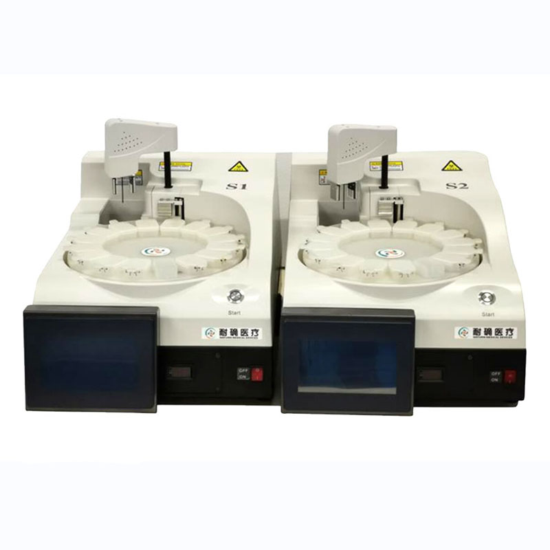
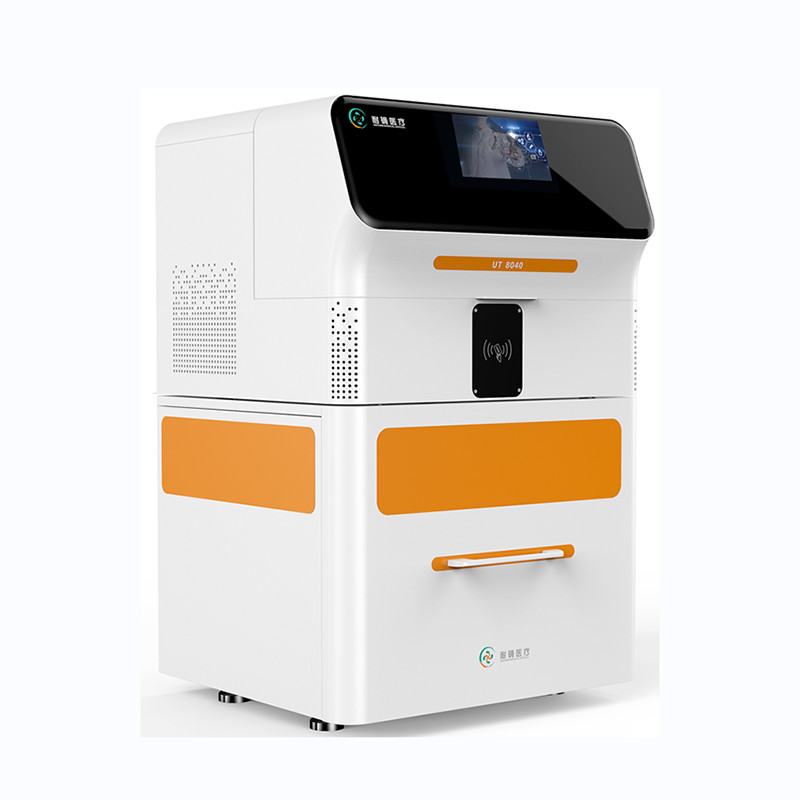
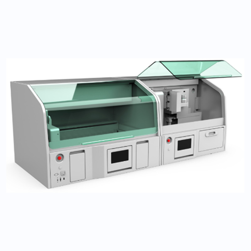
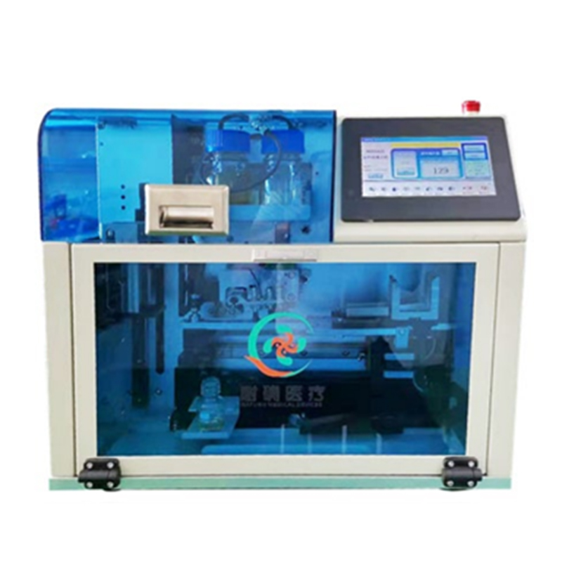
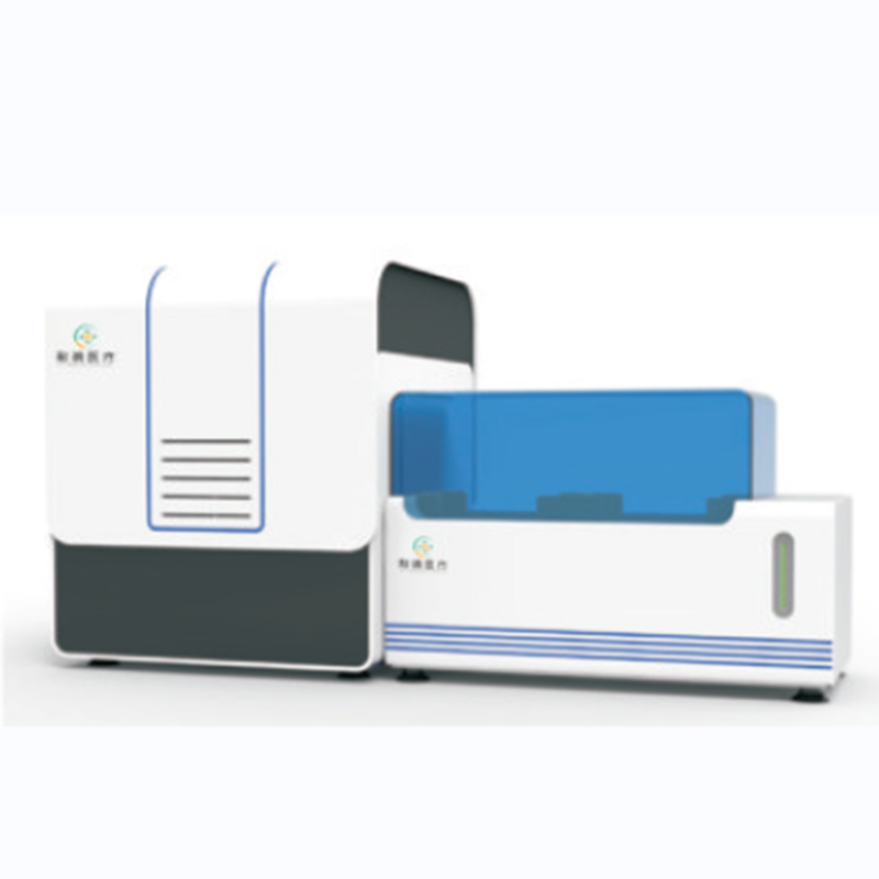
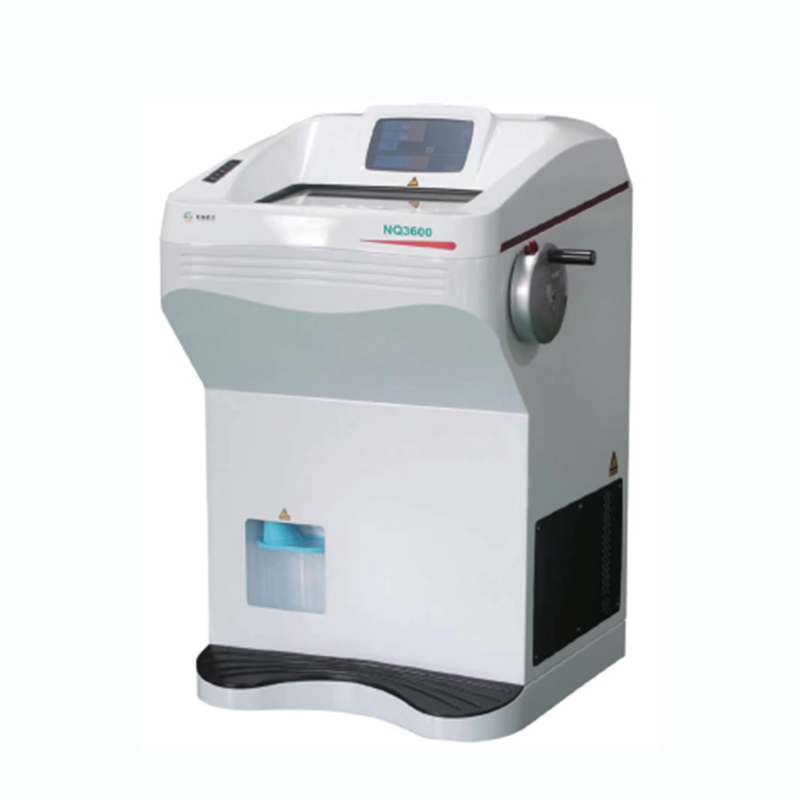
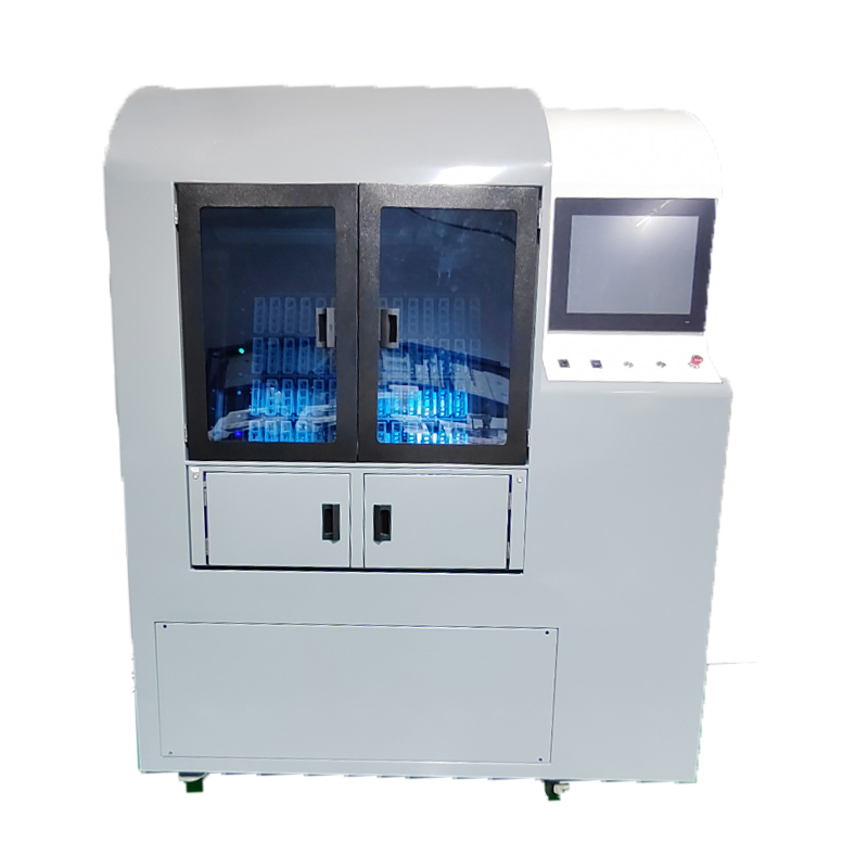
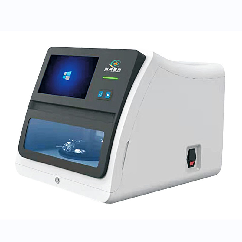
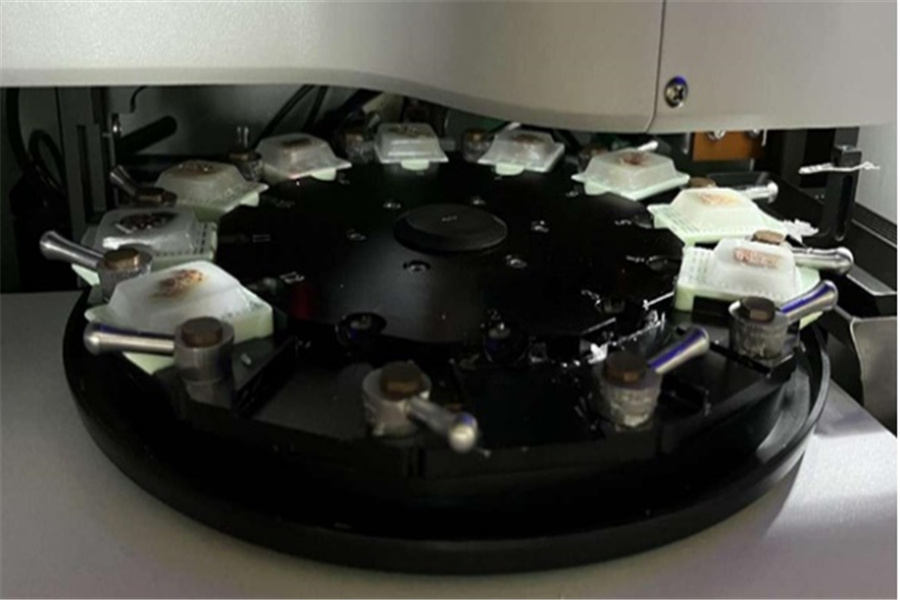
Automatic Tissue Microarrayer AUTO 12A

Automatic Tissue Microarrayer extracts tissues from region of interest on donor blocks and transplants them into the corresponding hole of recipient block automatically, creating perfect tissue microarrays for you, allowing scores of tissues to be analyzed on a single slide, valuable and efficient way for diagnostic and research purposes.

AUTO12A is a great technical breakthrough and of epoch-making significance, first in China, at the cutting edge of the world technology, with cost effectiveness.

    Video

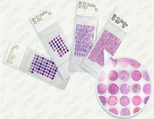
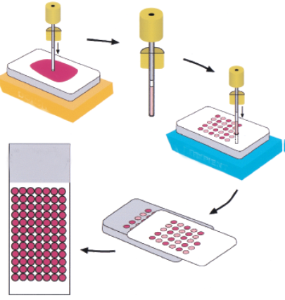
    Tissue Microarray Making Process

    1. Select the tissues to be studied.
    2. Mark the areas to be studied after HE staining.
    3. Use Tissue Microarrayer to extract the marked tissues per the user design, and implant to recipient paraffin block.
    4. After melting the paraffin, use microtome to section embedded tissues continuously to obtain tissue microarray.
    inguin6kc

    Features

    • 1. Realize automatic data entry and export the file.
    • 2. High cost effectiveness
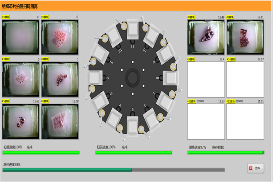
    • 3. Equipped with 5-megapixel autofocus camera and LED lighting to obtain real and clear images.
    • 4. PCR and biological sample management modules available for users to meet different experimental needs.
    The visual operation interface is clear, friendly and efficientwwq

    5. The visual operation interface is clear, friendly and efficient.

    The visual operation interface is clear, friendly and efficientms3

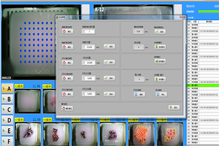
    6. Adopt patented precise positioning technology, with professional software which automatically punches the recipient block per the user design, extracts tissues from donor blocks, and makes the sampling needle move in X-axis and Y-axis directions, to make tissue microarray with the same bore diameter, implanting depth and spacing. 

    The visual operation interface is clear, friendly and efficientypf

    7. Take around 30 minutes to complete a 100-hole microarray, realizing unattended operation, autosuggesting after completion.

    The visual operation interface is clear, friendly and efficientmnx

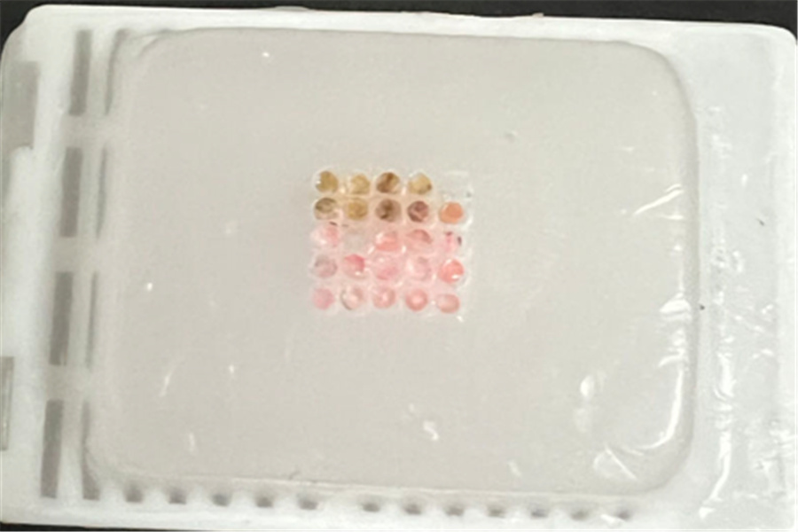
    8. The tissues matrix is neat and immune to any interference, with high precision.

    Specifications

    Block capacity

    67 (1 Recipient and 66 Donors)

    Core diameter

    0.6, 1.0, 1.5, 2.0, 2.5, 3.0 mm

    Speed

    200-250 cores/hour

    Hole precision

    ±0.001 mm

    Loading way

    Rotary disc

    Program

    NQDesigner

    Imaging

    5-megapixel high-resolution camera with autofocus

    Lighting

    LED

    Identification

    QR code, Barcode reader

    Optional modules

    PCR, Biological Sample Management

    Dimension

    560x520x510 mm

    Weight

    43 kg

    Applications

    Tissue microarray (TMA) is a new type of biochip technology, also known as Tissue Microarray Technology. The original intention was to do parallel research on large numbers of tissue samples in one experiment. 
    At present, it has been widely applied in the field of molecular biology, such as human gene research, protein research, drug development, biobanks, pathological daily quality control, AI training, and daily research of central laboratory research groups etc.
    asadfs5qc
    1. Using tissue chips for immunohistochemistry and special staining quality control
    2. Combined application with spatial omicsasdasedwetjm
    3. Combined application with fluorescence in situ hybridization (FISH)asdf10onn